What is Cartilage Defect?
A cartilage defect is a damaged area in the smooth surface inside the knee.
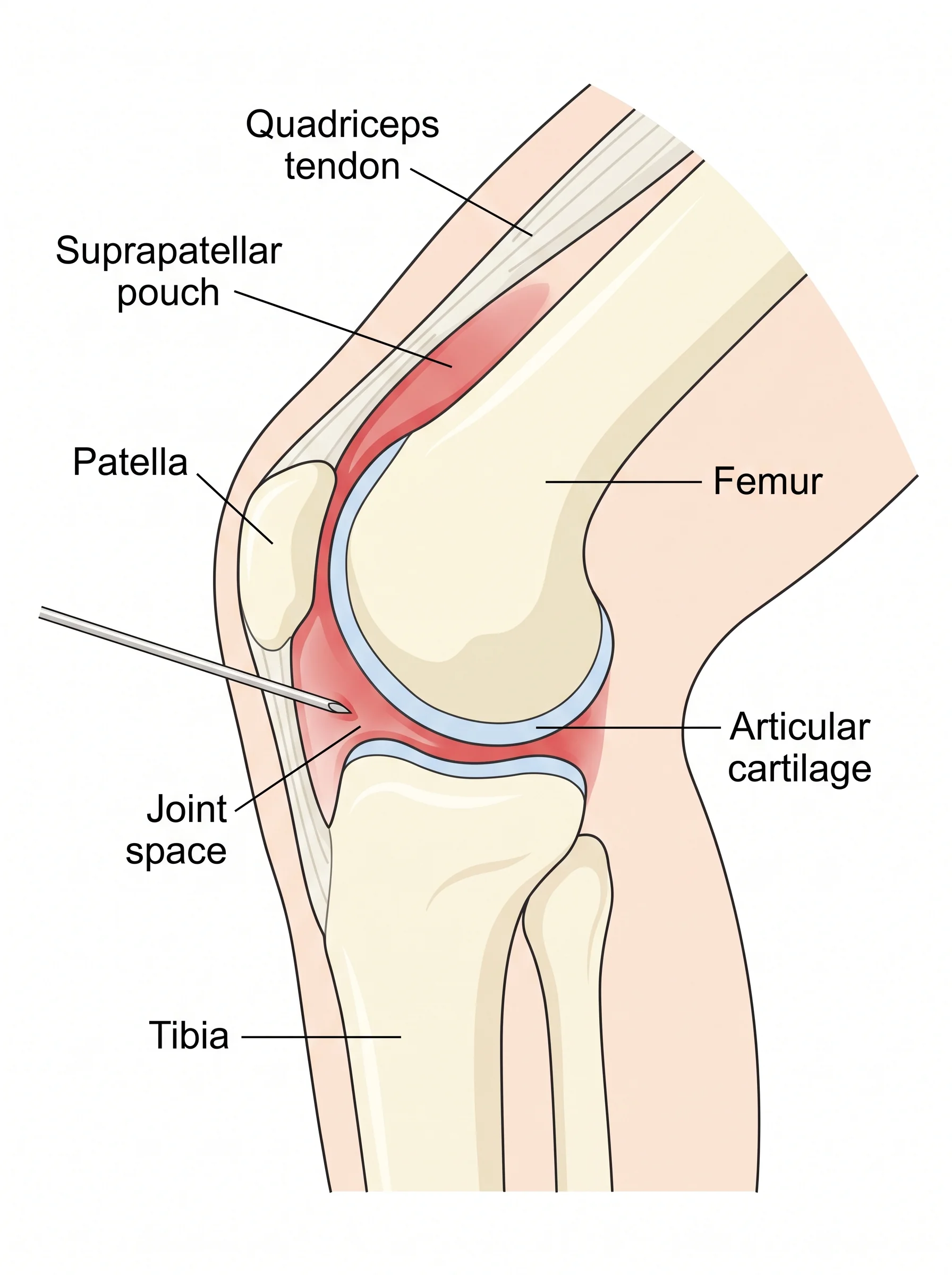
This surface, called articular cartilage, caps the ends of the bones so they glide against each other without pain. Unlike the meniscus, which is a separate cushion, articular cartilage is the slippery coating on the bone surfaces themselves. A defect is a spot where this coating is worn, cracked, or missing, sometimes down to the bone beneath.
Cartilage defects arise in a few ways. Some follow an injury, such as a twist or impact that shears off a piece of the surface, sometimes along with a ligament injury. Others develop gradually as part of early joint wear. A specific type, where a small area of bone and its cartilage lose blood supply and can loosen, is called osteochondritis dissecans, which tends to affect younger people.
An important feature of articular cartilage is that it has little to no blood supply and a limited ability to heal itself. A defect often does not simply fill back in on its own the way skin or bone might. This is why cartilage problems are approached thoughtfully, with treatment matched to the size, depth, and location of the defect and to the person's age and activity.
Defects range widely. A small, shallow spot may cause few symptoms and be managed simply, while a larger, deeper defect can cause ongoing pain and catching and may benefit from a procedure to repair or restore the surface. Because a defect can also lead to more widespread wear over time if left unaddressed, evaluation matters.
This deep guide pairs with the shorter cartilage defect service page at the office of Reza Jazayeri. Start there for a quick overview, then use this page for depth.
What are the symptoms of Cartilage Defect, and when should you see a specialist?
Cartilage defects can be quiet or troublesome, depending on their size and location.
Common experiences include the following.
- Pain in the knee, often felt with activity or when loading the affected area. - Swelling that comes and goes, sometimes after activity. - A catching, clicking, or locking feeling, especially if a fragment is loose. - A sense that something is moving inside the knee. - The knee giving way or feeling unreliable. - Stiffness or trouble with full motion.
When a piece of cartilage or bone has loosened, the knee may catch or lock as the fragment gets caught, which can be distinct and uncomfortable. When a defect is small and stable, symptoms may be mild and activity-related.
It is reasonable to see a specialist when:
- Knee pain and swelling persist beyond a few weeks without a clear explanation. - The knee catches, locks, or feels like something is loose inside. - Symptoms follow a twisting or impact injury. - Pain limits activity, work, or sport. - You have been told you may have a cartilage or osteochondritis dissecans problem.
Seek prompt care if the knee locks and will not move, if you cannot bear weight after an injury, or if there is significant swelling, since a loose fragment or combined injury may need earlier evaluation.
How is Cartilage Defect diagnosed, and what does the visit involve?
Diagnosing a cartilage defect relies on your history, a physical exam, and imaging, since cartilage is not seen on plain X-rays.
Your visit typically includes:
- A history. How symptoms began, whether there was an injury, and whether the knee catches, locks, or swells. - A physical exam. The specialist checks range of motion, presses to find tender areas, tests the ligaments and meniscus, and looks for signs of a loose fragment or swelling. - X-rays. These show the bones and joint space and can reveal a piece of bone that has loosened, as in osteochondritis dissecans, though they do not show the cartilage surface itself. - MRI. This magnet-based scan shows the cartilage in detail, revealing the size, depth, and location of a defect and whether the bone beneath is involved. It also shows companion injuries to the meniscus or ligaments.
Mapping the defect precisely is essential, because treatment depends heavily on how large and deep it is, where it sits, and whether the underlying bone is affected. The specialist combines the imaging with your symptoms and activity level to build a plan that fits both the defect and your goals.
What is the natural course, and what is the procedure sequence?
The course of a cartilage defect depends on its size, depth, location, and cause.
Small, stable defects may stay quiet, causing only occasional activity-related symptoms, and can often be managed without surgery for a long time. Because cartilage has little capacity to heal, the defect itself may not fill in, but with muscle support and load management the knee can feel and function well.
Larger or deeper defects, or those with a loose fragment, are more likely to cause ongoing symptoms such as catching, swelling, and pain. Left unaddressed, a significant defect can add stress to the surrounding surface and contribute to broader wear over time, which is one reason active people with symptomatic defects are evaluated carefully.
When surgery is chosen, the specific steps depend on the technique, most of which use arthroscopy, a small camera and thin instruments through tiny incisions. In general sequence, a cartilage procedure involves:
- Anesthesia, given so you feel no pain during surgery, discussed with the anesthesia team beforehand. - Inspection of the joint on a screen to confirm the defect's size, depth, and location and to address any loose fragments. - Preparing the defect, cleaning its edges and the base so any repair or new tissue can take hold. - The restorative step, which varies by technique. This may mean stabilizing or fixing a fragment, stimulating the underlying bone to form new surface tissue, or transplanting or growing cartilage to fill the defect. Some larger procedures are staged or use small incisions beyond arthroscopy. - Closure of the incisions.
Because these techniques aim to build or protect a living surface, the rehabilitation afterward is deliberate and often protective early, giving the repair time before full loading. The surgeon selects the approach that fits your defect and explains what recovery will involve.
What is the recovery timeline for Cartilage Defect?
Recovery from a cartilage defect varies widely with the treatment, because restorative procedures need time before full loading.
The milestones below are based on what you can do, alongside general guidance from the American Academy of Orthopaedic Surgeons. These are ranges, not promises.
Non-surgical management. The goal is to reduce symptoms and build supporting strength so the knee handles activity comfortably. With activity adjustment and guided exercise, many people improve meaningfully over about six to twelve weeks. The milestone is comfortable walking and low-impact activity without frequent swelling or catching.
After a cartilage procedure. Recovery is staged and often protective early to let the repair take hold. Limited weight bearing and a careful, gradual return of motion are common in the first weeks. The goal in this phase is to protect the new or repaired surface while keeping the joint moving gently. Rebuilding strength and returning to higher-impact activity is often staged over several months, and for some restorative techniques the full timeline extends toward a year. The milestone is meeting your surgeon's healing and strength targets before advancing load, not reaching a set date.
Across all paths, protecting the cartilage surface early is what allows durable function later. Progress is judged by healing and function rather than the calendar alone, and following the staged plan is central to a good result.
How should you prepare for Cartilage Defect?
If a cartilage procedure is chosen, planning ahead helps recovery.
Whether it is same-day surgery depends on the technique, which your team will confirm.
Prehabilitation. Before surgery, work to reduce swelling, keep motion, and maintain thigh strength. A calmer, stronger knee going in supports a smoother recovery.
Home setup. Because early weight bearing may be limited, set up a comfortable resting spot with room to elevate the leg, clear walkways, and keep daily items within easy reach. Prepare for using crutches for a period.
Transportation. Plan for someone to drive you home after anesthesia and to follow-up visits, since you may be limited in weight bearing for a while.
Time off. Discuss time away from work or school based on your job and the procedure. Restorative cartilage techniques often need more protected recovery than simpler procedures.
Support at hand. Have someone available for the first days, and possibly longer, to help at home while your weight bearing is limited.
Equipment. Have crutches, any prescribed brace, and ice therapy ready before surgery. Ask whether a motion device or specific aids are recommended for your technique.
Health preparation. Follow instructions on eating, drinking, and medications, and tell the team about all medicines and supplements. The office can answer insurance questions directly.
Which warning signs should you call about?
During recovery, call the office if you notice the following.
- Increasing redness, warmth, or drainage from an incision, or a fever, which can signal infection. - Swelling or pain that worsens instead of improving. - The knee locking or catching, which can suggest a loose fragment. - Calf pain, swelling, warmth, or tenderness, which should be checked promptly. - Numbness, tingling, or a cold, pale foot. - Bearing weight beyond what you were told to, or pain not controlled by your plan.
For severe symptoms such as sudden chest pain, trouble breathing, or a large, hot, swollen leg, seek emergency care right away. For other questions, call the office of Reza Jazayeri at (844) 529-0911; the team is glad to help you tell normal recovery from a concern, especially since early protection matters for cartilage.
What results can you expect from Cartilage Defect?
People understandably want to know how cartilage treatment turns out.
Results vary widely with the size, depth, and location of the defect, the technique, and the person, and the figures below are ranges, not promises.
According to general guidance from the American Academy of Orthopaedic Surgeons, small or less symptomatic cartilage defects can often be managed without surgery, with activity adjustment and strengthening controlling symptoms for many people.
General guidance from the American Academy of Orthopaedic Surgeons also notes that cartilage restoration procedures can relieve symptoms and improve function for appropriately selected people, that results depend heavily on the size and location of the defect and on following a staged rehabilitation, and that recovery for some techniques extends over many months.
The overall message is that many cartilage defects can be managed successfully, whether without surgery or with a procedure matched to the defect, and that careful selection and patient rehabilitation are central to a good result. The specialist will interpret these general figures in the context of your knee.
What are the treatment options?
- 01Activity adjustmentEasing off high-impact loading of the affected area gives symptoms a chance to settle. Low-impact activity that keeps the joint moving without aggravating it is encouraged.
- 02Guided exerciseA physical therapist, a clinician who teaches targeted exercises, strengthens the muscles around the knee and hip to share load and support the joint, and restores motion. Strong, balanced muscles can reduce symptoms and protect the surface.
- 03Pain reliefIce and, when appropriate, anti-inflammatory medicine can ease pain and swelling. Discuss regular use with your clinician.
- 04Weight supportBecause cartilage carries load, reducing extra body weight lowers the force across the defect and can ease symptoms, according to general guidance from the American Academy of Orthopaedic Surgeons.
- 05InjectionsIn some cases, an injection into the joint can reduce inflammation and support a return to exercise.
- 06When surgery is consideredSurgery is considered for defects that stay symptomatic despite non-surgical care, for loose fragments that catch or lock the knee, and for larger or deeper defects in active people where restoring the surface may protect the joint. Several procedures exist, matched to the defect. These range from smoothing or stabilizing the area, to techniques that encourage new surface tissue to form, to procedures that transplant or grow cartilage to fill a defect. If a fragment can be fixed back in place, that may be possible. The right choice depends on the size, depth, and location of the defect and on your age and activity, and the specialist explains the options so the decision is shared.
What do patients ask most?
Will a cartilage defect heal on its own?+
Is a cartilage defect the same as arthritis?+
How do you decide which treatment I need?+
Why is recovery from cartilage surgery so gradual?+
Can I stay active with a cartilage defect?+
Education and training
Medical school at Albert Einstein College of Medicine. Residency at NYU Hospital for Joint Diseases. Fellowship at Kerlan-Jobe Orthopaedic Clinic.
- FellowshipKerlan-Jobe Orthopaedic ClinicSports medicine
- ResidencyNYU Hospital for Joint DiseasesOrthopedic surgery residency
- Medical schoolAlbert Einstein College of MedicineDoctor of Medicine
- Board statusAmerican Board of Orthopaedic SurgeryBoard certified
DOCS Health
DOCS Satellite Westlake
696 Hampshire Rd
Suite 100 / Suite 180, Westlake Village, CA 91361
Phone(844) 529-0911